General Overview
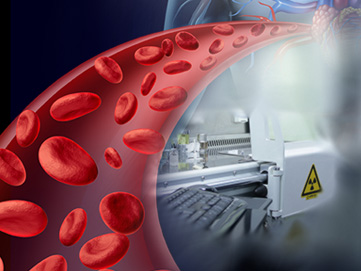
 Radionuclide angiography (RNA) is also known as a multi-gated acquisition (MUGA) scan, is a nuclear imaging test to help the doctor understand how the patient’s heart pumps blood. Radionuclide is a radioactive tracer. A special camera is used to take pictures of the heart as it pumps blood.
MUGA scan compares the heart’s function at rest. The physician may suggest the scan if an earlier ECG or EKG shows a problem and if the echo-cardiogram wasn’t helpful in determining heart function.
How It’s Done
The technician places small electrodes on the patient’s chest, arms and legs. The electrodes are connected to an electrocardiograph machine. Information about the patient’s heart is recorded. The ECG tracks the heartbeat. An intravenous line (IV) is used to inject the radionuclide. There is typically no pain involved, but the patient may experience a cool sensation as the radionuclide travels in the blood stream. The patient lies down on a table to rest. A special camera takes pictures of the heart and blood flow during the rest. What methods are used: A variety of methods are used to determine the patient’s heart function, including:- Echo-cardiogram
- Myocardial perfusion scintigraphy imaging
- Cardiac single photon emission computed tomography
- Radionuclide ventriculography – MUGA
- Magnetic resonance imaging
- Computed tomography
Any Side Effects?
RNA is a radioactive substance. The small amount used in the test prevents body injury. The patient’s kidneys process and excrete RNA within 24 hours. Pregnant women should not use RNA. Radiation can harm the unborn child. People with contrast dye allergies should tell the doctor before the procedure. Follow-up treatments or next steps: The test procedure allows you to soon resume normal activities. Immediately after the procedure, you are advised to drink plenty of water to remove the radioactive material from body. If you notice any pain or swelling on the spot where the IV line was introduced, notify the doctor immediately.
